


At EmergeOrtho—Triangle Region, our orthopedic specialists often rely on Magnetic Resonance Imaging (MRI) tests to diagnose musculoskeletal conditions that affect the neck. An MRI of the cervical spine can identify injuries and conditions in the soft tissues and bones, allowing providers to reach a faster diagnosis and start treatment to get you back to doing the things you love even sooner.
If you have been scheduled for an MRI of the cervical spine, we want you to know what to expect. Understanding why you are having the test, what your doctor is looking for, and how to prepare will help you feel more comfortable throughout the entire process.
What is a Cervical Spine MRI and Why Would You Need One?
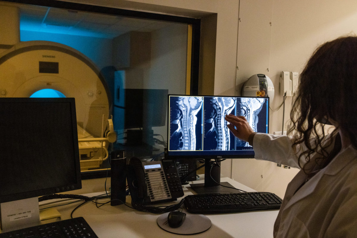
An MRI is a diagnostic imaging test that uses a magnetic field and computer-generated radio waves — not radiation, like X-rays — to produce images of the soft tissues, organs, and bones inside your body. It’s a noninvasive test that produces detailed, high-resolution, cross-sectional images called slices. Every exam produces multiple slices, allowing the doctor to view multiple angles and get a complete picture of what is happening inside your body.
When you have an MRI of the cervical spine, the images focus on the upper part of your spine and neck. The cervical spine has seven small bones (vertebrae), which start at the base of the skull and run to the base of the neck. Your neck also has more than 20 muscles, which are connected to the spine via tendons. These provide support, help you move your head around, and assist with basic functions like chewing, swallowing, and breathing.
The most common reason doctors order a cervical spine MRI is to diagnose severe neck, shoulder, or arm pain that doesn’t improve with treatment. MRIs are also ordered for patients who have neck pain accompanied by weakness or numbness in the arms and legs.
Other reasons to schedule an MRI of the cervical spine include:
- Assessing injury or trauma to the spine after an accident
- Evaluating arthritis progression
- Evaluating spinal birth defects
- Identifying infections that involve the spine
- Treating severe scoliosis (spinal curvature)
- Treating multiple sclerosis
- Identifying a spinal tumor
Physicians may schedule a cervical spine MRI before neck surgery, or when other imaging tests do not provide adequate results.
What Doctors are Looking for from MRI Images of the Cervical Spine
When you have an MRI of the cervical spine, “normal” results mean there are no obvious issues with the vertebrae, spinal discs, or surrounding soft tissues and nerves.
An abnormal result means that the test revealed an issue within your cervical spine. Some of the most common abnormal results include:
- Cervical radiculopathy, or a herniated spinal disc
- Spinal stenosis, a narrowing of the spinal column
- Cervical spondylosis, or excessive wear on the neck bones and cartilage
- Age-related degenerative conditions (including arthritis)
- Discitis, or disc inflammation
Other, less common, abnormal test results include bone or spinal column infections, spinal fracture, tumors, and spinal cord injuries.
Depending on the results of the MRI of the cervical spine, your doctor will recommend a treatment plan, which may include everything from non-invasive treatments (a large category of treatments including physical therapy and medication) to surgery.
How to Prepare for an MRI of the Cervical Spine
Because of the powerful magnets used in the MRI, some patients may not be able to safely undergo the test. Before your appointment, talk to your doctor and let them know if you have certain metal or electronic devices in your body that could be a safety hazard or affect image quality. These devices include:
- Implantable heart defibrillators, pacemakers, drug infusion pumps, or nerve stimulators
- Metal artificial joints
- Metal stents, pins, screws, rods, or plates
- Cochlear implants
- Intrauterine device (IUD)
- Metal fragments of any type
- Certain tattoos or artificial makeup, which may contain metal
Some of these devices are “MRI safe,” but you need to discuss them with the doctor. You should also let your doctor know if you are pregnant or breastfeeding.
During the Test
During the MRI, the Technologist may inject a contrast dye into your bloodstream to help improve image quality.
You will lie on a table, which is moved into a tube that is open on either end. Some people feel claustrophobic, and can take anti-anxiety medication to help them relax. It may also be possible to have an “open MRI,” which has a larger scanning area.
During the test, you need to lie still to prevent blurred images, but you will not feel anything as the radio waves move around your body. The machine will make noise, but you can block it out with earplugs, or even listen to music to help you relax.
Once the MRI is complete, you can resume normal activities while you wait for the results.
Cervical Spine MRI at EmergeOrtho
When you have a cervical spine MRI at EmergeOrtho—Triangle Region, you can expect the highest quality of care at any of our convenient locations. Our innovative MRI services allow the team of board-certified orthopedic surgeons to make fast, accurate diagnoses for a wide range of conditions. In fact, our imaging services have achieved the gold standard in medical imaging with national recognition for excellence through accreditation from the American College of Radiology (ACR).
If neck pain is disrupting your life, self-schedule an appointment with one of our providers today, and start on the journey to Emerge Stronger. Healthier. Better. You can also call us any time with questions at 984.279.3649.


